Publications
Selected publications by categories in reversed chronological order (full list can be found in my CV). * denotes equal contributions.
2025
-
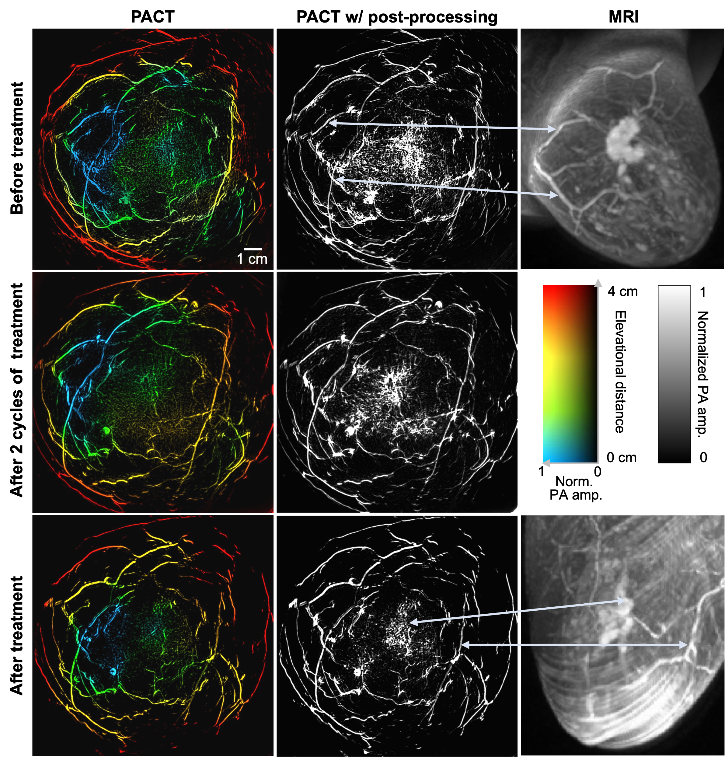
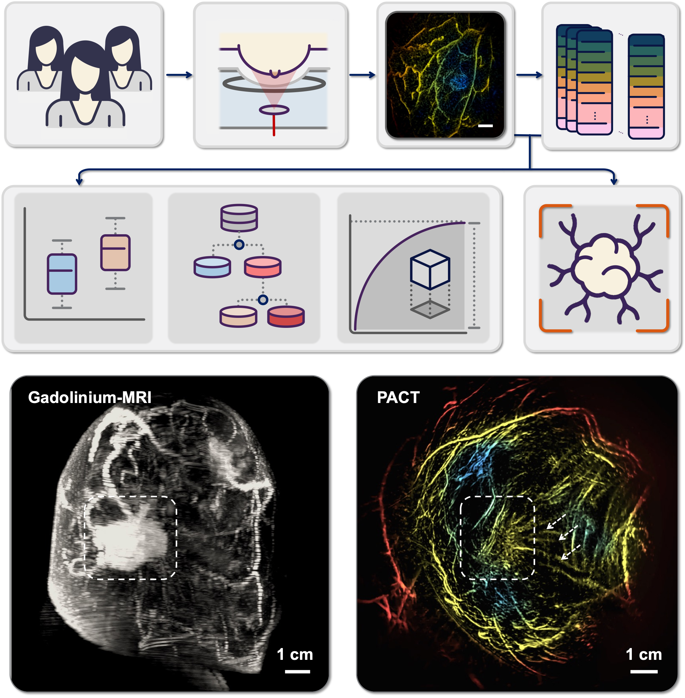 Panoramic photoacoustic computed tomography with learning-based classification enhances breast lesion characterizationXin Tong*, Cindy Z Liu*, Yilin Luo*, and 8 more authorsNature Biomedical Engineering, 2025
Breast cancer diagnosis is crucial due to the high prevalence and mortality rate associated with the disease. However, mammography involves ionizing radiation and has compromised sensitivity in radiographically dense breasts, ultrasonography lacks specificity and has operator-dependent image quality, and magnetic resonance imaging faces high cost and patient exclusion. Photoacoustic computed tomography (PACT) offers a promising solution by combining light and ultrasound for high-resolution imaging that detects tumour-related vasculature changes. Here we introduce a workflow using panoramic PACT for breast lesion characterization, offering detailed visualization of vasculature irrespective of breast density. Analysing PACT features of 78 breasts in 39 patients, we develop learning-based classifiers to distinguish between normal and suspicious tissue, achieving a maximum area under the receiver operating characteristic curve of 0.89, which is comparable with that of conventional imaging standards. We further differentiate malignant and benign lesions using 13 features. Finally, we developed a learning-based model to segment breast lesions. Our study identifies PACT as a non-invasive and sensitive imaging tool for breast lesion evaluation.
2024
-
 Quantum imaging of biological organisms through spatial and polarization entanglementYide Zhang*, Zhe He*, Xin Tong*, and 3 more authorsScience Advances, 2024
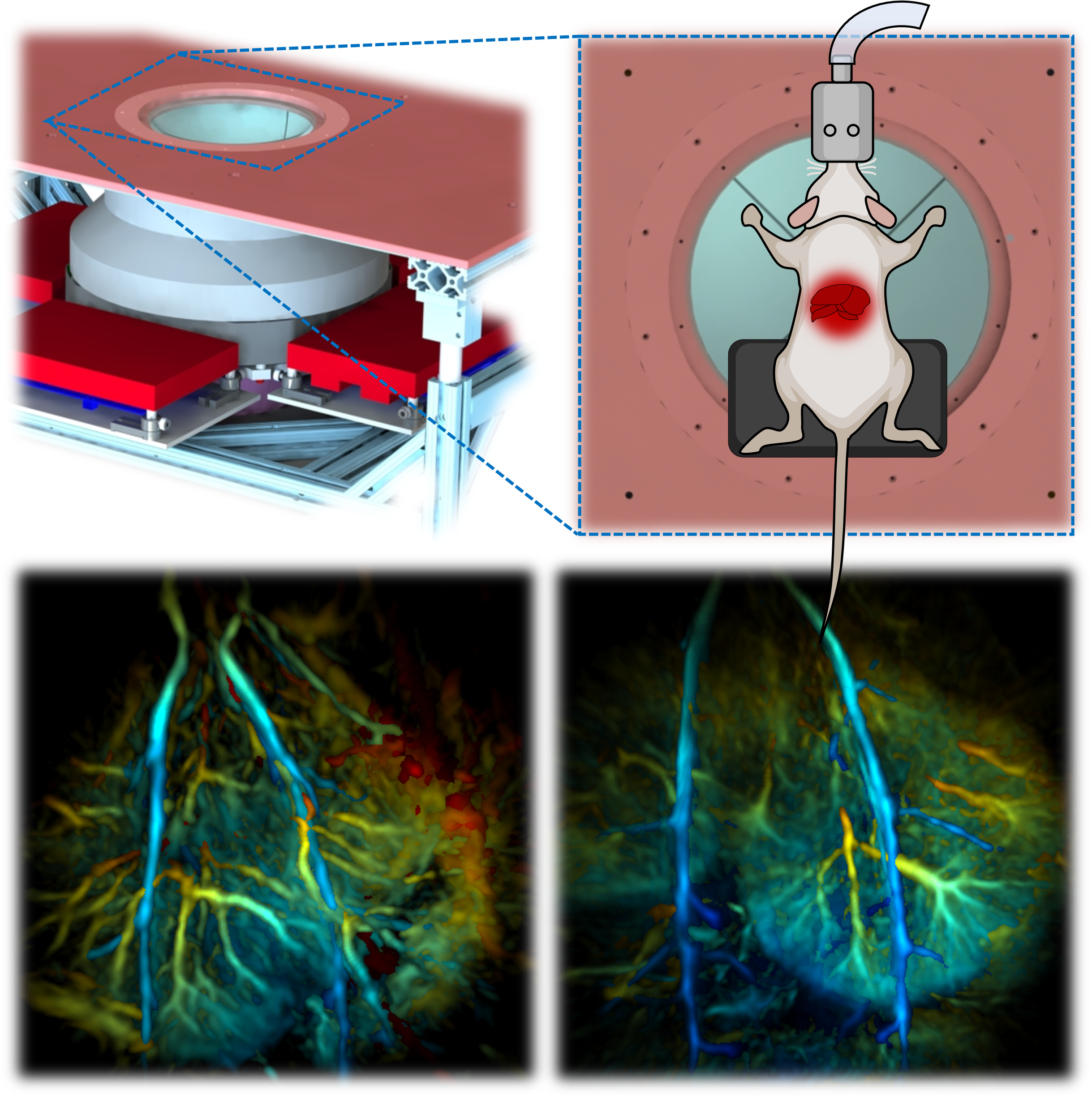
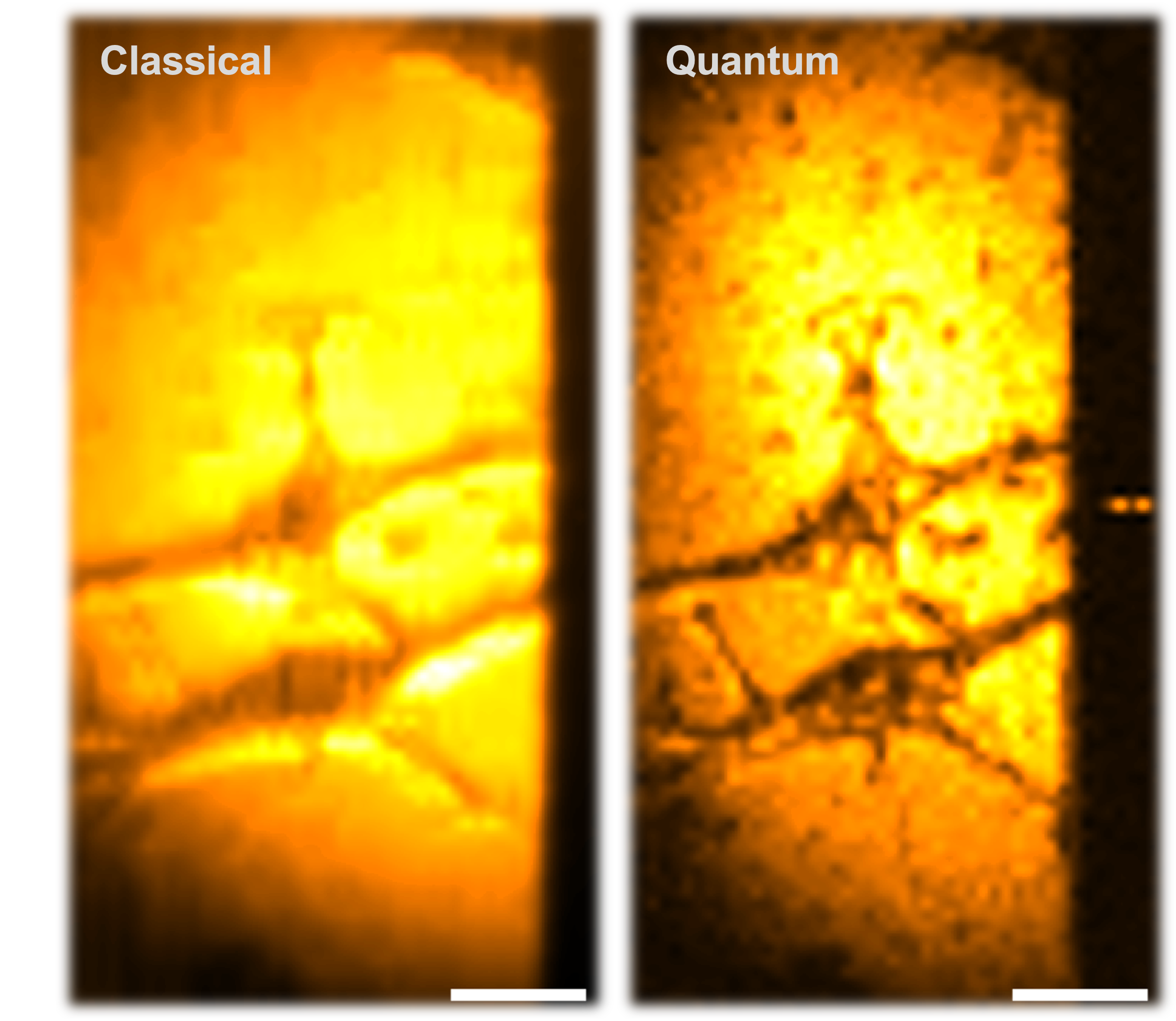
Quantum imaging holds potential benefits over classical imaging but has faced challenges such as poor signal-to-noise ratios, low resolvable pixel counts, difficulty in imaging biological organisms, and inability to quantify full birefringence properties. Here, we introduce quantum imaging by coincidence from entanglement (ICE), using spatially and polarization-entangled photon pairs to overcome these challenges. With spatial entanglement, ICE offers higher signal-to-noise ratios, greater resolvable pixel counts, and the ability to image biological organisms. With polarization entanglement, ICE provides quantitative quantum birefringence imaging capability, where both the phase retardation and the principal refractive index axis angle of an object can be remotely and instantly quantified without changing the polarization states of the photons incident on the object. Furthermore, ICE enables 25 times greater suppression of stray light than classical imaging. ICE has the potential to pave the way for quantum imaging in diverse fields, such as life sciences and remote sensing.
-
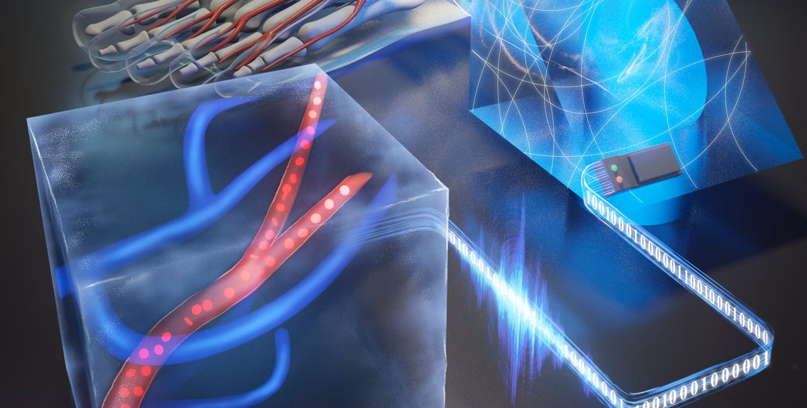 Ultrafast longitudinal imaging of haemodynamics via single-shot volumetric photoacoustic tomography with a single-element detectorYide Zhang*, Peng Hu*, Lei Li, and 8 more authorsNature biomedical engineering, 2024
Techniques for imaging haemodynamics use ionizing radiation or contrast agents or are limited by imaging depth (within approximately 1 mm), complex and expensive data-acquisition systems, or low imaging speeds, system complexity or cost. Here we show that ultrafast volumetric photoacoustic imaging of haemodynamics in the human body at up to 1 kHz can be achieved using a single laser pulse and a single element functioning as 6,400 virtual detectors. The technique, which does not require recalibration for different objects or during long-term operation, enables the longitudinal volumetric imaging of haemodynamics in vasculature a few millimetres below the skin’s surface. We demonstrate this technique in vessels in the feet of healthy human volunteers by capturing haemodynamic changes in response to vascular occlusion. Single-shot volumetric photoacoustic imaging using a single-element detector may facilitate the early detection and monitoring of peripheral vascular diseases and may be advantageous for use in biometrics and point-of-care testing.
-
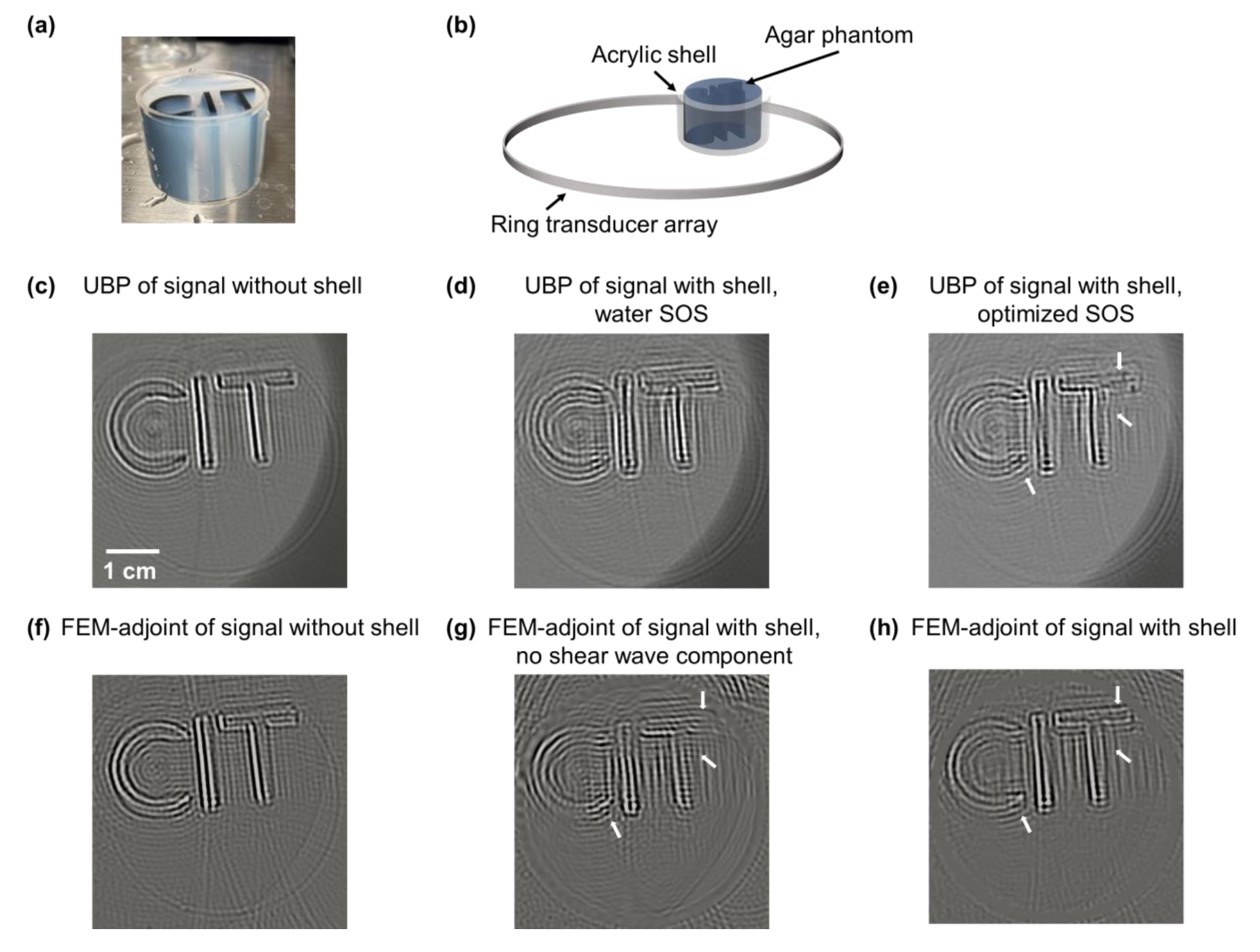 Full-wave Image Reconstruction in Transcranial Photoacoustic Computed Tomography using a Finite Element MethodYilin Luo, Hsuan-Kai Huang, Karteekeya Sastry, and 8 more authorsIEEE transactions on medical imaging, 2024
Transcranial photoacoustic computed tomography presents challenges in human brain imaging due to skull-induced acoustic aberration. Existing full-wave image reconstruction methods rely on a unified elastic wave equation for skull shear and longitudinal wave propagation, therefore demanding substantial computational resources. We propose an efficient discrete imaging model based on finite element discretization. The elastic wave equation for solids is solely applied to the hard-tissue skull region, while the soft-tissue or coupling-medium region that dominates the simulation domain is modeled with the simpler acoustic wave equation for liquids. The solid-liquid interfaces are explicitly modeled with elastic-acoustic coupling. Furthermore, finite element discretization allows coarser, irregular meshes to conform to object geometry. These factors significantly reduce the linear system size by 20 times to facilitate accurate whole-brain simulations with improved speed. We derive a matched forward-adjoint operator pair based on the model to enable integration with various optimization algorithms. We validate the reconstruction framework through numerical simulations and phantom experiments.
-
 Enhancing Optical Microscopy with Quantum EntanglementXin Tong, Yide Zhang, and Lihong V. WangOptics & Photonics News, 2024
Drawing on the unique properties of entangled photons, quantum approaches can overcome the limitations of classical methods, enhance spatial resolution and reduce stray light and shot noise.
2023
-
 Non-Invasive 3D Photoacoustic Tomography of Angiographic Anatomy and Hemodynamics of Fatty Livers in RatsXin Tong*, Li Lin*, Peng Hu, and 4 more authorsAdvanced Science, 2023
Non-alcoholic fatty liver disease is the most common liver disorder worldwide, which strongly correlates to obesity, diabetes, and metabolic syndromes. Complementary to mainstream liver diagnostic modalities, photoacoustic tomography (PAT) can provide high-speed images with functional optical contrast. However, PAT has not been demonstrated to study fatty liver anatomy with clear volumetric vasculatures. The livers of multiple rats are non-invasively imaged in vivo using the recently developed 3D PAT platform. The system provides isotropically high spatial resolution in 3D space, presenting clear angiographic structures of rat livers without injecting contrast agents. Furthermore, to quantitatively analyze the difference between the livers of lean and obese rats, the authors measured several PAT features and statistical differences between the two groups are observed. In addition to the anatomy, a time-gated strategy is applied to correct respiration-induced motion artifacts and extracted the hemodynamics of major blood vessels during the breathing cycles. This study demonstrates the capabilities of 3D-PAT to reveal both angiographic anatomy and function in rat livers, providing hematogenous information for fatty liver diagnosis. 3D-PAT, as a new tool for preclinical research, warrants further improvements to be transferred to human pediatric liver imaging.
-
 Non-invasive photoacoustic computed tomography of rat heart anatomy and functionLi Lin*, Xin Tong*, Susana Cavallero, and 5 more authorsLight: Science & Applications, 2023
Complementary to mainstream cardiac imaging modalities for preclinical research, photoacoustic computed tomography (PACT) can provide functional optical contrast with high imaging speed and resolution. However, PACT has not been demonstrated to reveal the dynamics of whole cardiac anatomy or vascular system without surgical procedure (thoracotomy) for tissue penetration. Here, we achieved non-invasive imaging of rat hearts using the recently developed three-dimensional PACT (3D-PACT) platform, demonstrating the regulated illumination and detection schemes to reduce the effects of optical attenuation and acoustic distortion through the chest wall; thereby, enabling unimpeded visualization of the cardiac anatomy and intracardiac hemodynamics following rapidly scanning the heart within 10 s. We further applied 3D-PACT to reveal distinct cardiac structural and functional changes among the healthy, hypertensive, and obese rats, with optical contrast to uncover differences in cardiac chamber size, wall thickness, and hemodynamics. Accordingly, 3D-PACT provides high imaging speed and nonionizing penetration to capture the whole heart for diagnosing the animal models, holding promises for clinical translation to cardiac imaging of human neonates.
-
 Quantum microscopy of cells at the Heisenberg limitZhe He*, Yide Zhang*, Xin Tong*, and 2 more authorsNature Communications, 2023
Entangled biphoton sources exhibit nonclassical characteristics and have been applied to imaging techniques such as ghost imaging, quantum holography, and quantum optical coherence tomography. The development of wide-field quantum imaging to date has been hindered by low spatial resolutions, speeds, and contrast-to-noise ratios (CNRs). Here, we present quantum microscopy by coincidence (QMC) with balanced pathlengths, which enables super-resolution imaging at the Heisenberg limit with substantially higher speeds and CNRs than existing wide-field quantum imaging methods. QMC benefits from a configuration with balanced pathlengths, where a pair of entangled photons traversing symmetric paths with balanced optical pathlengths in two arms behave like a single photon with half the wavelength, leading to a two-fold resolution improvement. Concurrently, QMC resists stray light up to 155 times stronger than classical signals. The low intensity and entanglement features of biphotons in QMC promise nondestructive bioimaging. QMC advances quantum imaging to the microscopic level with significant improvements in speed and CNR toward the bioimaging of cancer cells. We experimentally and theoretically prove that the configuration with balanced pathlengths illuminates an avenue for quantum-enhanced coincidence imaging at the Heisenberg limit.
-
 Experimental full-domain mapping of quantum correlation in Clauser-Horne-Shimony-Holt scenariosXin Tong*, Zhe He*, Yide Zhang*, and 4 more authorsPhysical Review Applied, 2023
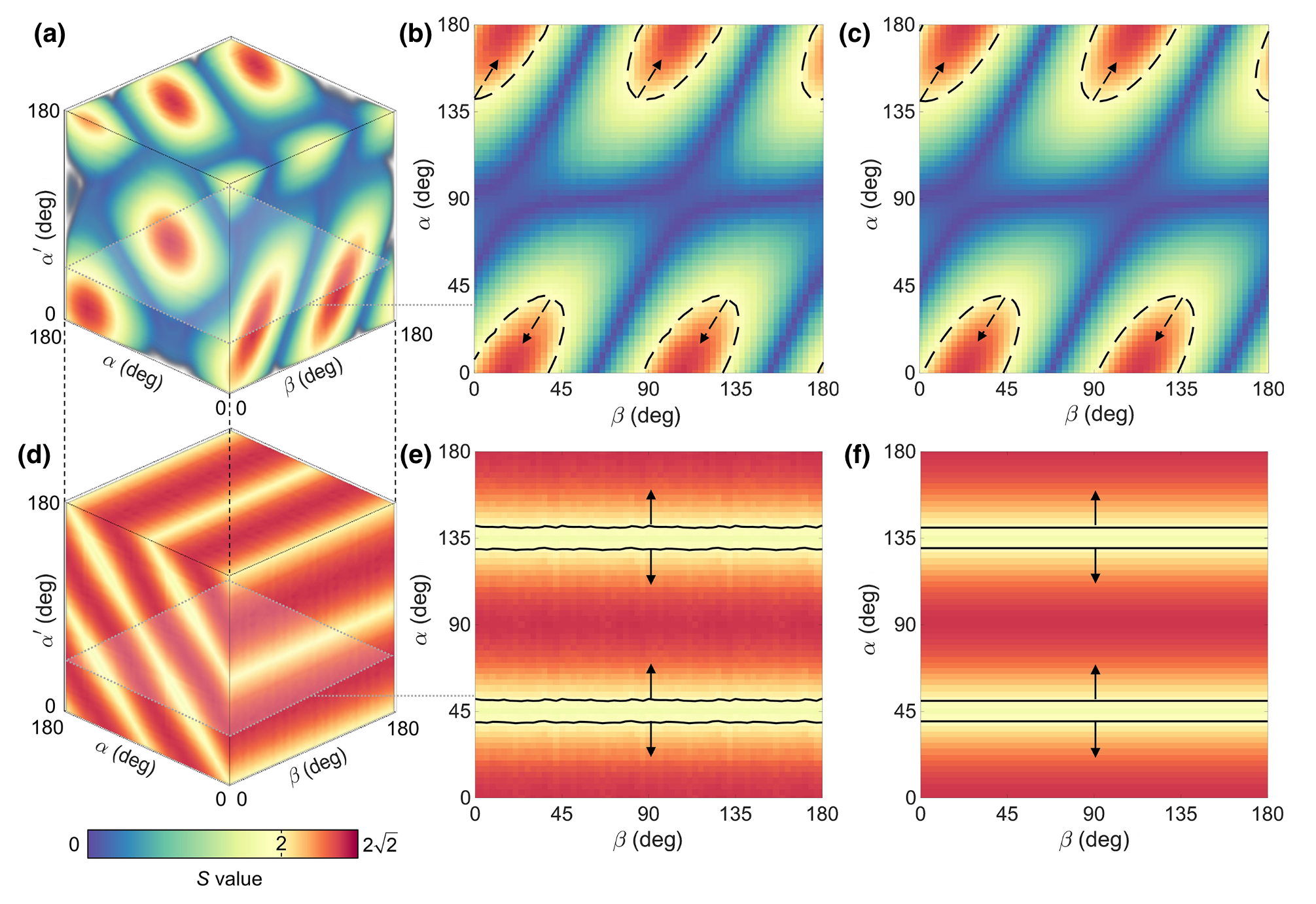
Quantum correlation between two parties serves as a useful resource in the surging applications of quantum information. The Bell nonlocality and quantum steering have been proposed to describe nonclassical correlations against local-hidden-variable and local-hidden-state theories, respectively. To characterize the two types of nonclassical correlations, various nonlocality and steering inequalities have been established, and the amount of inequality violation serves as a helpful indicator for many entanglement-based tasks. Quantum state tomography has been employed for measuring quantum states, while the method requires intensive computation and does not directly verify either nonlocality or steering over the full domain independent of established theories. Here, we experimentally map the full-domain correlation with bipartite states for nonlocality and quantum steering in Clauser-Horne-Shimony-Holt scenarios. The measurement of the maps automatically accounts for detection imperfections. Furthermore, we demonstrate the application of the correlation maps in the entanglement-based quantum key distribution protocol with arbitrary bipartite states. The correlation maps show direct measurements and simple interpretations that can answer fundamental questions about nonlocality and quantum steering as well as contribute to quantum information applications in a straightforward manner.
-
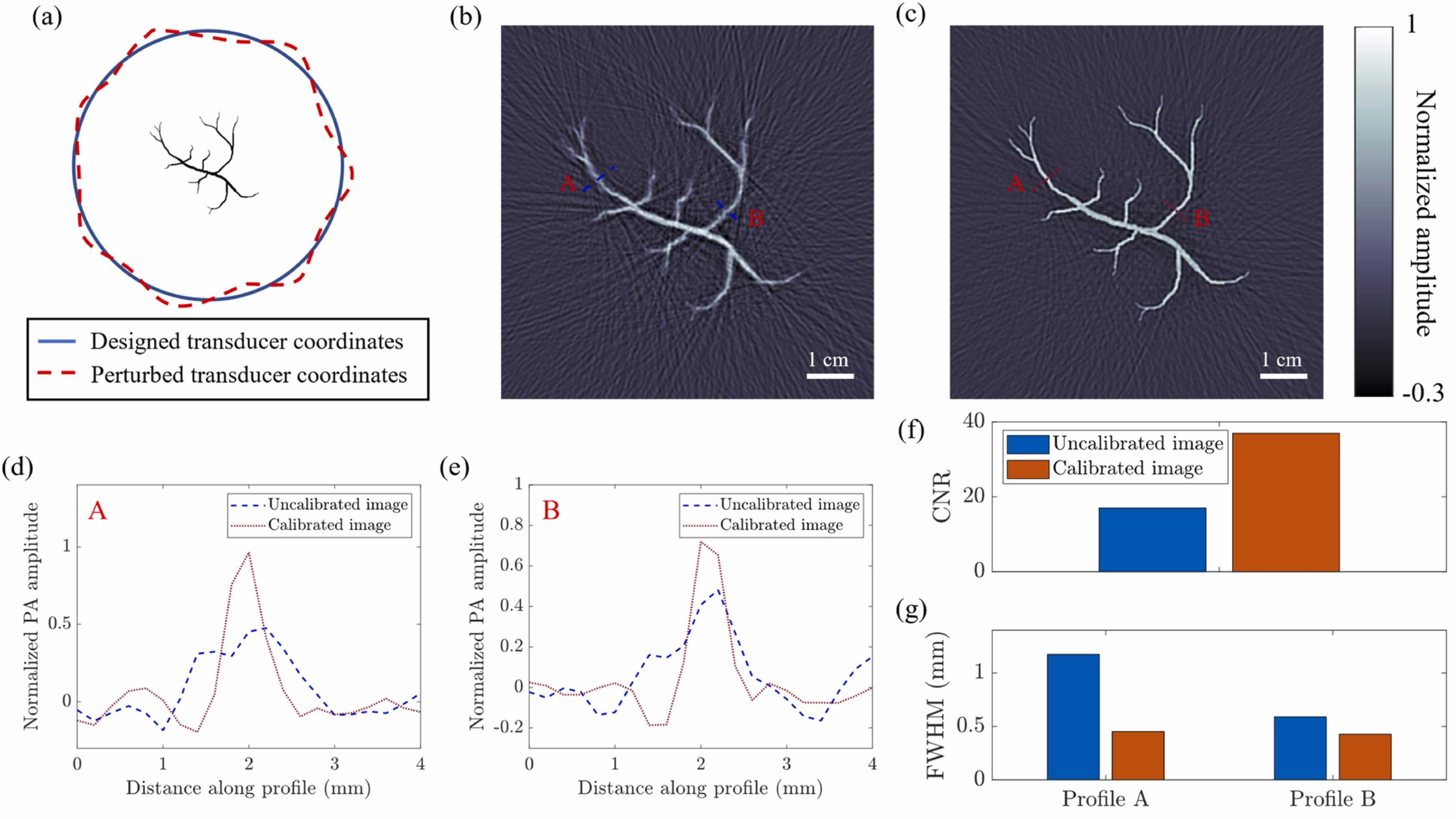 A method for the geometric calibration of ultrasound transducer arrays with arbitrary geometriesKarteekeya Sastry, Yang Zhang, Peng Hu, and 4 more authorsPhotoacoustics, 2023
Geometric calibration of ultrasound transducer arrays is critical to optimizing the performance of photoacoustic computed tomography (PACT) systems. We present a geometric calibration method that is applicable to a wide range of PACT systems. We obtain the speed of sound and point source locations using surrogate methods, which results in a linear problem in the transducer coordinates. We characterize the estimation error, which informs our choice of the point source arrangement. We demonstrate our method in a three-dimensional PACT system and show that our method improves the contrast-to-noise ratio, the size, and the spread of point source reconstructions by 80±19%, 19±3%, and 7±1%, respectively. We reconstruct the images of a healthy human breast before and after calibration and find that the calibrated image reveals vasculatures that were previously invisible. Our work introduces a method for geometric calibration in PACT and paves the way for improving PACT image quality.
2021
-
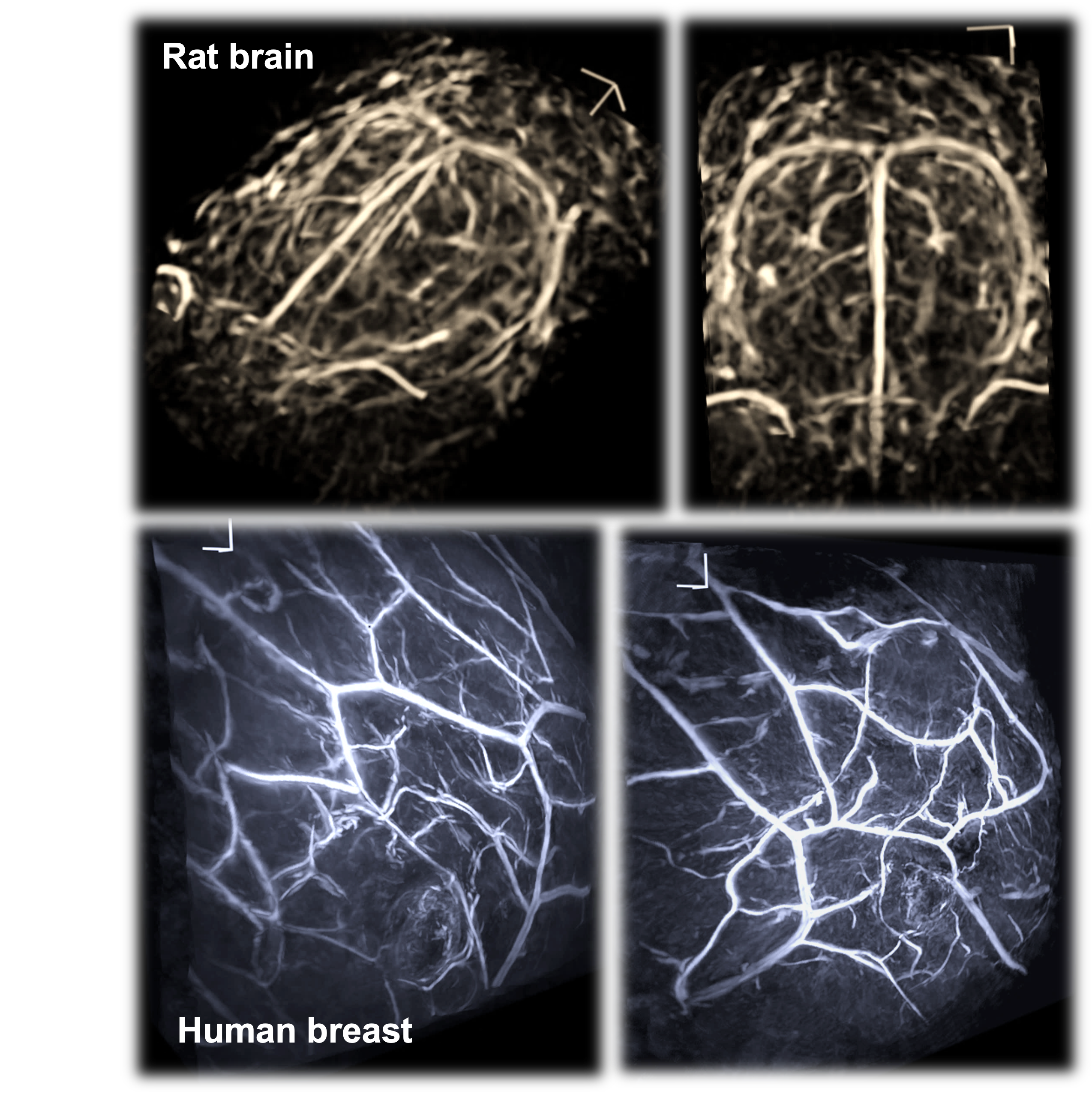 High-speed three-dimensional photoacoustic computed tomography for preclinical research and clinical translationLi Lin*, Peng Hu*, Xin Tong*, and 7 more authorsNature Communications, 2021
Photoacoustic computed tomography (PACT) has generated increasing interest for uses in preclinical research and clinical translation. However, the imaging depth, speed, and quality of existing PACT systems have previously limited the potential applications of this technology. To overcome these issues, we developed a three-dimensional photoacoustic computed tomography (3D-PACT) system that features large imaging depth, scalable field of view with isotropic spatial resolution, high imaging speed, and superior image quality. 3D-PACT allows for multipurpose imaging to reveal detailed angiographic information in biological tissues ranging from the rodent brain to the human breast. In the rat brain, we visualize whole brain vasculatures and hemodynamics. In the human breast, an in vivo imaging depth of 4 cm is achieved by scanning the breast within a single breath hold of 10 s. Here, we introduce the 3D-PACT system to provide a unique tool for preclinical research and an appealing prototype for clinical translation.
-
 Photoacoustic computed tomography of breast cancer in response to neoadjuvant chemotherapyLi Lin*, Xin Tong*, Peng Hu, and 3 more authorsAdvanced Science, 2021
Neoadjuvant chemotherapy (NAC) has contributed to improving breast cancer outcomes, and it would ideally reduce the need for definitive breast surgery in patients who have no residual cancer after NAC treatment. However, there is no reliable noninvasive imaging modality accepted as the routine method to assess response to NAC. Because of the inability to detect complete response, post-NAC surgery remains the standard of care. To overcome this limitation, a single-breath-hold photoacoustic computed tomography (SBH-PACT) system is developed to provide contrast similar to that of contrast-enhanced magnetic resonance imaging, but with much higher spatial and temporal resolution and without injection of contrast chemicals. SBH-PACT images breast cancer patients at three time points: before, during, and after NAC. The analysis of tumor size, blood vascular density, and irregularity in the distribution and morphology of the blood vessels on SBH-PACT accurately identifies response to NAC as confirmed by the histopathological diagnosis. SBH-PACT shows its near-term potential as a diagnostic tool for assessing breast cancer response to systemic treatment by noninvasively measuring the changes in cancer-associated angiogenesis. Further development of SBH-PACT may also enable serial imaging, rather than the use of current invasive biopsies, to diagnose and follow indeterminate breast lesions.
2018
-
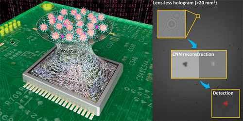 Deep learning enables high-throughput analysis of particle-aggregation-based biosensors imaged using holographyYichen Wu, Aniruddha Ray, Qingshan Wei, and 5 more authorsACS Photonics, 2018
Aggregation-based assays, using micro- and nanoparticles have been widely accepted as an efficient and cost-effective biosensing tool, particularly in microbiology, where particle clustering events are used as a metric to infer the presence of a specific target analyte and quantify its concentration. Here, we present a sensitive and automated readout method for aggregation-based assays using a wide-field lens-free on-chip microscope, with the ability to rapidly analyze and quantify microscopic particle aggregation events in 3D, using deep learning-based holographic image reconstruction. In this method, the computation time for hologram reconstruction and particle autofocusing steps remains constant, regardless of the number of particles/clusters within the 3D sample volume, which provides a major throughput advantage, brought by deep learning-based image reconstruction. As a proof of concept, we demonstrate rapid detection of herpes simplex virus (HSV) by monitoring the clustering of antibody-coated microparticles, achieving a detection limit of ∼5 viral copies/μL (i.e., ∼25 copies/test).
2017
-
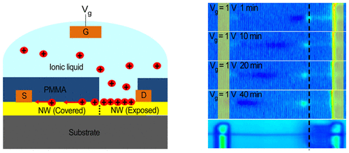 In situ visualization of fast surface ion diffusion in vanadium dioxide nanowiresYasen Hou, Rui Xiao, Xin Tong, and 2 more authorsNano letters, 2017
We investigate in situ ion diffusion in vanadium dioxide (VO2) nanowires (NWs) by using photocurrent imaging. Alkali metal ions are injected into a NW segment via ionic liquid gating and are shown to diffuse along the NW axis. The visualization of ion diffusion is realized by spatially resolved photocurrent measurements, which detect the charge carrier density change associated with the ion incorporation. Diffusion constants are determined to be on the order of 10–10 cm2/s for both Li+ and Na+ ions at room temperature, while H+ diffuses much slower. The ion diffusion is also found to occur mainly at the surface of the NWs, as metal contacts can effectively block the ion diffusion. This novel method of visualizing ion distribution is expected to be applied to study ion diffusion in a broad range of materials, providing key insights on phase transition electronics and energy storage applications.